Cone Beam 3D Imaging in Hickory, NC
At Hickory Dentistry in Hickory, NC, we use Cone Beam 3D imaging to see what traditional X-rays simply can’t show — giving Dr. Millsaps a complete, three-dimensional view of your teeth, roots, bone, airways, and sinuses in under ten seconds. This technology means faster diagnoses, more precise treatment planning, and better outcomes for our patients. Call (828) 322-2977 to schedule your appointment today.
Advanced Cone Beam 3D Imaging at Hickory Dentistry in Hickory, NC
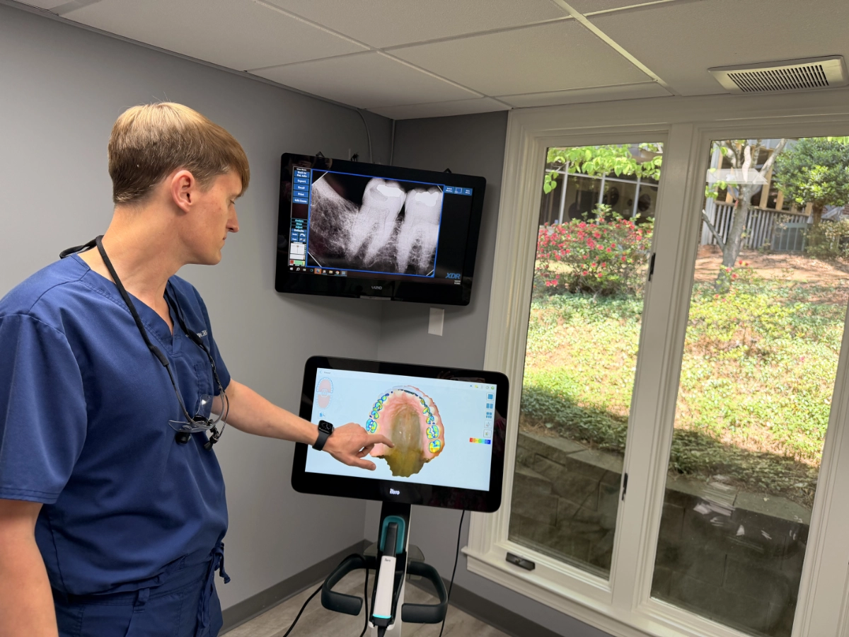
Accurate diagnosis is the foundation of great dental care — and Cone Beam Computed Tomography (CBCT) imaging represents one of the most significant advancements in diagnostic dentistry available today. Unlike conventional two-dimensional X-rays, which provide a flat, limited view of your oral structures, our Cone Beam 3D imaging system captures a complete, detailed three-dimensional picture of your teeth, roots, jawbone, nerves, airways, sinuses, and temporomandibular joints in a single, rapid scan. The entire process takes less than ten seconds and the images are available for review almost instantly — allowing Dr. Millsaps to begin discussing findings and treatment options with you during the same appointment.
At Hickory Dentistry in Hickory, NC , we invest in this technology because we believe every patient in Hickory, NC and the surrounding communities of Newton, Lenoir, Granite Falls, and Conover deserves the most accurate diagnosis modern dentistry can provide. Cone Beam imaging helps us catch problems sooner, plan treatments more precisely, and reduce the number of surprises — for both our team and our patients — during complex procedures.
How Cone Beam 3D Imaging Works
The Cone Beam CT scanner rotates around your head in a single pass, projecting a cone-shaped beam of X-ray radiation that captures hundreds of images from multiple angles simultaneously. These images are then processed by the imaging software to construct a highly accurate, three-dimensional digital model of your oral and facial structures. The resulting 3D dataset can be viewed as cross-sections from any angle, in any plane — giving Dr. Millsaps the ability to virtually “fly through” your anatomy and examine specific areas with unprecedented clarity. The radiation dose from a Cone Beam scan is significantly lower than a traditional medical CT scan and uses a fraction of the radiation — making it a safe and practical option for detailed diagnostic imaging. The entire scan takes less than ten seconds, the images are displayed within moments, and there is no discomfort involved — patients simply stand or sit still while the scanner rotates around them.
Clinical Applications of Cone Beam 3D Imaging
Cone Beam 3D imaging enhances nearly every aspect of complex dental diagnosis and treatment planning. For dental implant placement, it is an indispensable tool — it allows Dr. Millsaps to precisely measure available bone volume and density, map the exact location of critical structures like the inferior alveolar nerve and maxillary sinuses, and determine the optimal implant size, angle, and position before surgery. This level of pre-surgical planning dramatically reduces procedural risk and improves the accuracy of implant placement. For patients requiring complex root canal therapy, 3D imaging reveals the full root canal anatomy — including extra canals and unusual root configurations that would be missed on flat X-rays — ensuring complete treatment. For patients being evaluated for sleep apnea, the airway view provided by CBCT imaging helps identify obstructions and informs oral appliance therapy planning. TMJ evaluation, wisdom tooth assessment, orthodontic treatment planning, and evaluation of cysts or pathology in the jaw are all enhanced significantly by the detail that 3D imaging provides. When Dr. Millsaps needs to see exactly what’s happening inside your jaw before making an important treatment decision, Cone Beam imaging is the tool that makes that precision possible.
Safety of Cone Beam 3D Imaging
At Hickory Dentistry, we understand that radiation exposure is a concern many patients have — and we take that concern seriously. Our Cone Beam CT system is calibrated to use the minimum effective radiation dose necessary to obtain a diagnostically useful image, and the dose is significantly lower than a conventional medical CT scan. We apply CBCT imaging selectively — only when the detailed three-dimensional information it provides is genuinely necessary for diagnosis or treatment planning that cannot be adequately accomplished with conventional digital X-rays alone. We follow the established ALARA principle (As Low As Reasonably Achievable) for all imaging decisions in our office. If you have specific questions about radiation exposure from dental imaging, our team is happy to discuss the specifics with you at any appointment.
Why Choose Hickory Dentistry for Advanced Dental Imaging in Hickory, NC?
Investing in Cone Beam 3D imaging technology is an investment in patient outcomes — and at Hickory Dentistry, we make that investment because our patients deserve the highest standard of diagnostic care available. The ability to see your complete oral anatomy in three dimensions gives Dr. Millsaps a level of diagnostic confidence that simply isn’t possible with flat X-rays alone, and that confidence translates directly into more accurate diagnoses, more precise treatments, and fewer complications. Patients from Hickory, NC and surrounding communities trust Hickory Dentistry because we consistently bring together advanced technology, clinical expertise, and genuine commitment to patient wellbeing. Call (828) 322-2977 or request an appointment online to experience the difference comprehensive diagnostic care makes.
Frequently Asked Questions — Cone Beam 3D Imaging in Hickory, NC
Is Cone Beam 3D imaging the same as a medical CT scan?
Cone Beam CT and medical CT both produce three-dimensional images, but they are different technologies designed for different purposes. Medical CT scanners use a fan-shaped beam and produce full-body cross-sectional images at significantly higher radiation doses. Cone Beam CT uses a focused, cone-shaped beam specifically designed for head and neck imaging in a dental setting, delivering detailed 3D images of the teeth, jaws, and surrounding structures at a much lower radiation dose than a medical CT. The Cone Beam scanner at Hickory Dentistry is calibrated to provide excellent diagnostic quality while minimizing patient exposure.
How long does a Cone Beam 3D scan take?
The actual scanning process takes less than ten seconds. You’ll simply sit or stand still while the scanner rotates around your head — there’s no discomfort, no injections, and nothing placed inside your mouth. The images are processed and available for review within minutes. In most cases, Dr. Millsaps can review the 3D dataset with you during the same appointment, discuss the findings, and begin planning your treatment without any delay. The entire imaging appointment is typically very brief and straightforward.
When is Cone Beam 3D imaging recommended versus standard X-rays?
Standard digital X-rays are appropriate for routine diagnosis — checking for cavities, monitoring bone levels, and evaluating teeth and their roots in most clinical situations. Cone Beam 3D imaging is recommended when the additional three-dimensional detail is genuinely necessary for accurate diagnosis or precise treatment planning. This most commonly includes dental implant planning, complex root canal cases, impacted tooth evaluation, TMJ assessment, sleep apnea airway evaluation, and investigation of jaw pathology. Dr. Millsaps follows evidence-based guidelines for imaging decisions and recommends CBCT only when the benefit clearly justifies it.
Does Cone Beam imaging hurt?
Not at all — Cone Beam 3D imaging is completely painless and non-invasive. No X-ray film is placed in your mouth, no needles are involved, and nothing touches your teeth or gums during the scan. You simply remain still for less than ten seconds while the scanner rotates. Patients who have experienced discomfort from traditional dental X-rays — particularly those with sensitive gag reflexes — often find the Cone Beam scan to be a much more comfortable diagnostic experience.
Can Cone Beam imaging be used to plan dental implants?
Yes — and in fact, 3D imaging is one of the most valuable tools available for dental implant planning. The detailed 3D dataset allows Dr. Millsaps to precisely measure the width, height, and density of available bone at the implant site, identify the exact position of critical anatomical structures like the inferior alveolar nerve and maxillary sinus, and determine the optimal implant size and placement angle with far greater confidence than is possible with flat X-rays. This pre-surgical precision reduces risk, improves accuracy, and contributes directly to better long-term implant outcomes. If you’re considering dental implants in Hickory, NC, Cone Beam imaging will be part of your evaluation.
Will my insurance cover Cone Beam 3D imaging?
Coverage for Cone Beam CT imaging varies by insurance plan. Many plans provide at least partial coverage when the imaging is associated with a covered procedure such as implant placement or surgical tooth extraction. Our team at Hickory Dentistry will verify your benefits before any imaging and provide a clear explanation of estimated costs so there are no surprises. If you have questions about coverage or out-of-pocket costs, please call (828) 322-2977 — we’re happy to help you understand your options.
Do you use Cone Beam 3D imaging for patients from Newton, Lenoir, and Granite Falls?
Hickory Dentistry provides Cone Beam 3D imaging and comprehensive dental technology services to patients from Hickory, NC and all surrounding communities, including Newton, Lenoir, Granite Falls, Conover, and beyond. If you’re looking for a dental practice that combines advanced diagnostic technology with personalized, expert care, we’d love to welcome you. Call (828) 322-2977 or request an appointment online today.